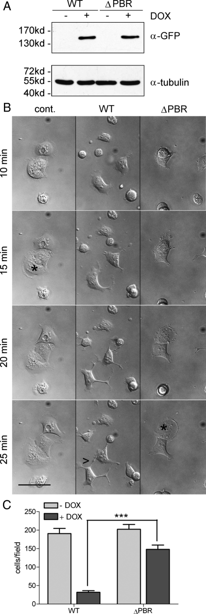
Figure 8.
A DLC1 mutant deficient in PI(4,5)P2 binding fails to inhibit cell spreading and directed migration. (A) HEK293 Flp-In DLC1 WT and ΔPBR cells were left untreated (−) or treated (+) with 100 ng/ml doxycycline overnight to induce DLC1 expression. Whole cell extracts were separated by SDS-PAGE and analyzed by Western blotting with GFP-specific (top) and tubulin-specific. antibodies (bottom) as a loading control. (B) HEK293 Flp-In DLC1 WT and ΔPBR cells were treated with 100 ng/ml doxycycline overnight. Noninduced HEK293 Flp-In DLC1 WT cells were used as a control. Cells were harvested and plated onto collagen-coated glass bottom dishes. After 5 min, bright-field images were taken every 30 s for 90 min. The figure shows snapshots of the movies (see Online Supplemental files) at the indicated time points; asterisks mark cells with lamellipodia; arrowheads point to projections. Scale bar, 50 μm. (C) HEK293 Flp-In DLC1 WT, and ΔPBR cells were left untreated or treated with 100 ng/ml doxycycline. Cells, 5 × 10⁴, were seeded in medium containing 0.5% FCS into the upper chamber of a Transwell. The lower well contained medium supplemented with 10% FCS. Cells that had migrated across the filter after 4 h were fixed and stained. The number of migrated cells was determined by counting five independent microscopic fields (20-fold magnification). Data shown are the mean of duplicate wells and are representative of two independent experiments. Error bars, SEM.

