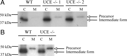
Figure 4.
UCE −/− mouse skin fibroblasts hypersecrete cathepsin D. (A) Primary skin fibroblasts isolated from wild-type or UCE −/− mice were incubated for 90 min with TRAN 35S-LABEL methionine/cysteine containing media (pulse) and then for 4 h in unlabeled media (chase) containing 5 mM of Man-6-P. Cathepsin D was immunoprecipitated from cell lysates (C) and media (M) collected at the end of the 4-h chase period and resolved on 10% SDS-PAGE. Signals were visualized by autoradiography. A representative experiment (of a total of 3 for WT and 6 for UCE −/−, with fibroblasts isolated from separate mice) is shown. Arrows indicate the proform of cathepsin D (∼47 kDa) and cathepsin D intermediate form (43 kDa). (B) Fibroblasts were incubated for 90 min with TRAN 35S-LABEL methionine/cysteine containing media and then for 4 h in unlabeled chase media containing 5 mM Man-6-P and 660 μg/ml HSA-mannose. Cathepsin D was immunoprecipitated as described in A (WT and UCE −/−, n = 3).

