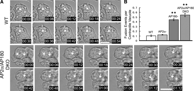
Figure 4.
Increased fusion of contractile vacuoles in the absence of AP180. (A) Time lapse of living wild-type cells and AP2α/AP180 DKO cells in water. In wild-type cells (WT), a whole life cycle of one contractile vacuole (arrow) is shown, from expansion to contraction. In AP2α/AP180 DKO cells (AP2α/AP180 DKO) two contractile vacuoles (arrows) fuse into a single contractile vacuole (arrow). Scale bar, 10 μm. See supplemental Movies S1 and S2 for the corresponding time lapse movies. (B) Quantification of homotypic fusion of contractile vacuoles in wild-type, AP2α−, AP180−, and AP2α/AP180 DKO cells. Error bar, SE, n = 3 independent experiments, 34 contractile vacuoles for each cell line quantified in each experiment. (*p <0.05, **p <0.005).

