Figure 10.

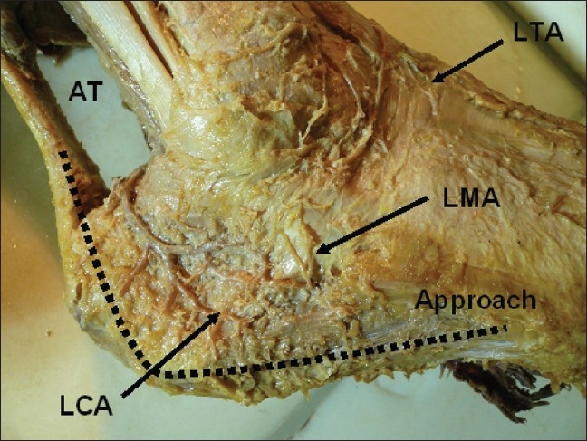
Cadaveric photograph shows soft tissue vascularization of the lateral wall of the hind foot on cadaver. LCA - lateral calcaneal artery, LTA - ventrolateral tarsal arteries, LMA - lateral malleolar artery, AT – Achilles tendon

Cadaveric photograph shows soft tissue vascularization of the lateral wall of the hind foot on cadaver. LCA - lateral calcaneal artery, LTA - ventrolateral tarsal arteries, LMA - lateral malleolar artery, AT – Achilles tendon