
Figure 1.
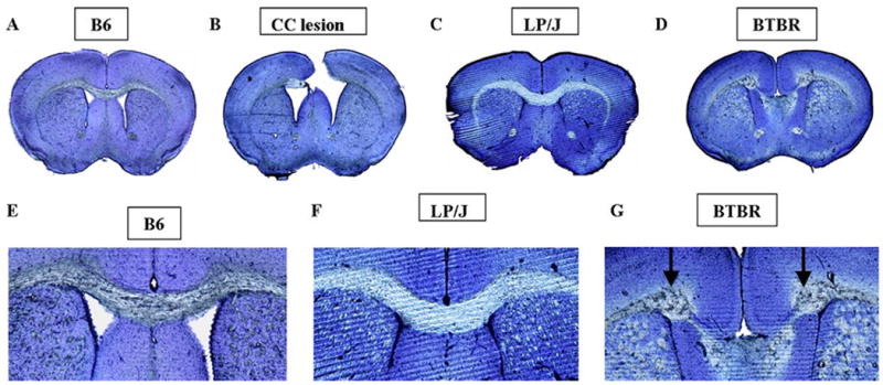
Photomicrographs of thioin-stained representative coronal sections depicting the corpus callosum (CC) region in representative (A) B6 control mice; (B) B6 mice in which the CC was lesioned on postnatal day 7 (tissue obtained from subjects sacrificed at 3 months of age); (C) LP/J mice with intact CC; (D) BTBR mice in which the corpus callosum is congenitally absent. Close-ups of the CC region are shown in (E) B6; (F) LP/J; (G) BTBR. Probst bundles appear in BTBR only (arrows). Scale bar 80=μm.