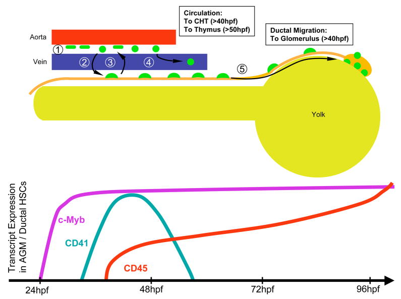
Figure 8.
Model of hematopoietic stem cell migration in the zebrafish embryo. (1) HSCs (green) appear between the dorsal aorta and cardinal vein. (2) Some HSCs translocate to the pronephric tubules (orange) and often back again to between the vessels (3). (4) Some HSCs enter circulation to seed the developing CHT and thymic anlage. (5) HSCs migrate anteriorly along each duct to generate the first hematopoietic cells in the developing kidney. Nascent HSCs can be visualized by expression of the c-myb, CD41, and CD45 genes (bottom panel).

