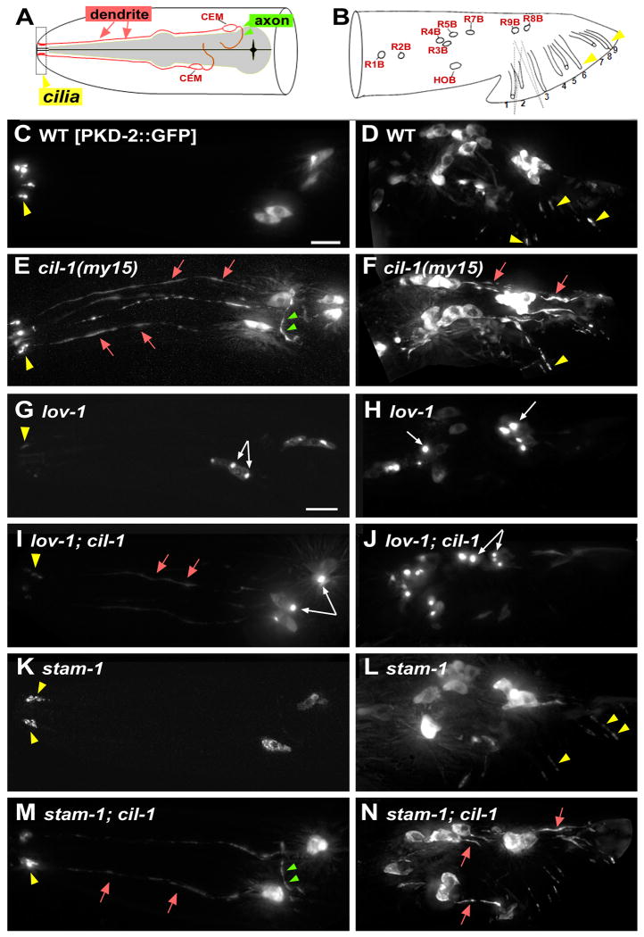
Figure 1. cil-1 is required for TRP polycystin complex (PKD-2 and LOV-1) localization. cil-1 acts between lov-1 and stam-1 in ray B neurons.
Cell bodies = white arrows, cilia = yellow arrowhead, dendrite = red arrow, axon = green arrowhead. (A-B) Cartoons illustrating locations and structure of pkd-2 expressing neurons in C. elegans male head (A) and tail (B). The head CEMs and tail ray neurons are bilateral, and only one side of the animal is shown. Modified from [37]. (C, D) In a WT male, PKD-2∷GFP localizes to cilia and neuronal cell bodies of CEM, ray B (RnB), and hook B (HOB) neurons. (E, F) In cil-1(my15) males, PKD-2∷GFP is abnormally distributed along neurons including dendrites and axons. PKD-2∷GFP in ciliary regions appears WT. (G) In lov-1 CEMs, PKD-2∷GFP accumulates in cell bodies and weakly labels in cilia. (H) In lov-1 RnBs, PKD-2∷GFP accumulates in cell bodies and is not detectable in cilia. (I) In cil-1; lov-1 CEMs, PKD-2∷GFP aggregates in cell bodies and distributes along dendrites and cilia. (J) In cil-1; lov-1 RnBs, PKD-2∷GFP forms bright aggregates in the cell bodies, similar to the lov-1 single mutant. (K) In stam-1 CEMs, PKD-2∷GFP accumulates in ciliary regions. (L) In stam-1 RnBs, PKD-2∷GFP accumulates in the ciliary regions and distal dendrites [15]. (M) In stam-1; cil-1 CEMs, PKD-2∷GFP localizes to dendrites and axons, and sometimes accumulates ciliary bases. (N) In stam-1; cil-1 RnBs, PKD-2∷GFP is distributed to dendritic and axonal processes, similar to cil-1 single mutants Scale bar, 10um.

