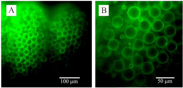
Figure 2.
Fluorophore-conjugated lectin binding to the cnidocyst. Fluorescence micrographs of Physalia tentacles stained with FITC-conjugated lectins. (A). Lower power image of part of a tentacle stained with FITC-conjugated isolectin B4 lectin. The two clusters of stained cysts represent two cnidosacs. (B). A higher power image of cysts stained with soybean agglutinin. Two size classes of cysts are evident in this image.

