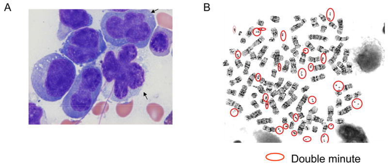
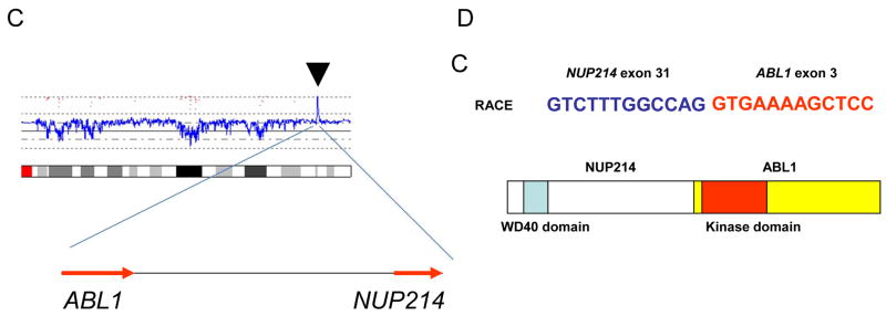
Fig. 1. NUP214-ABL1 gene in T-cell leukemic cells.

A: Morphology of leukemic cells. Atypical cells show convoluted nuclear contour, dense chromatin, and bizarre mitotic figures. Cells with “flower shape” nuclei are also observed (arrows).
B: Metaphase spreads. Double minute chromosomes are circled.
C: High copy number amplification of NUP214 and ABL1. The result of SNP-chip analysis (chromosome 9) revealed high copy number amplification of chromosome 9q34 region (arrow head) which contained NUP214 and ABL1 genes (arrows indicate the direction of transcription of the genes).
D: NUP214-ABL1 fusion was detected in T-cell leukemia. Rapid amplification of cDNA ends (RACE) method and nucleotide sequencing showed the fusion of exon 31 of NUP214 and exon 3 of ABL1. Schematic representation of the structure of NUP214-ABL1 fusion protein is shown.

