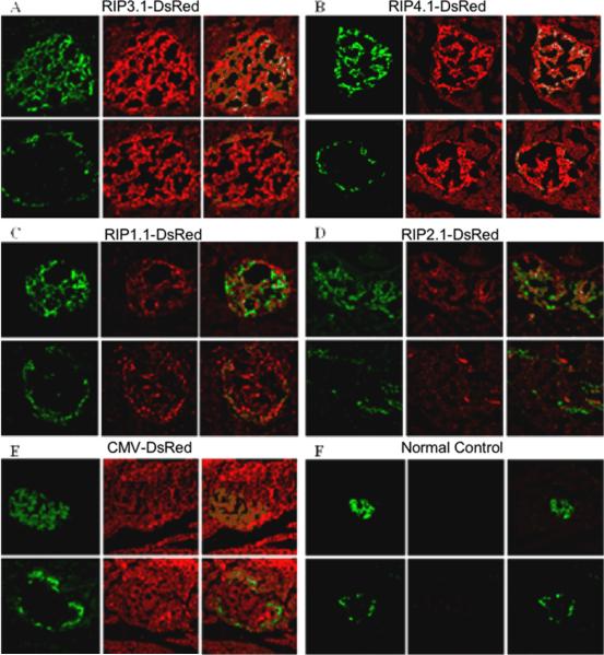
Figure 6.
In vivo immunohistology of rats treated with DsRed reporter gene using different RIP promoter lengths and controls. All images are magnified at 200X. For each group labeled A–F, a representative islet section is shown. Top panels are stained with green anti-insulin (left), red anti-DsRed (middle), and their confocal image (right). Bottom panels are adjacent sections of the same islet stained with green anti-glucagon (left), red anti-DsRed (middle), and their confocal image (right). A: RIP3.1-DsRed; B: RIP-4.1-DsRed; C: RIP-1.1-DsRed; D: RIP-2.1-DsRed slides, E: pCMV-DsRed; F: normal control.

