
Figure 7. Therapeutic stability and plasmid derived gene expression require continuous IL-10 signaling.
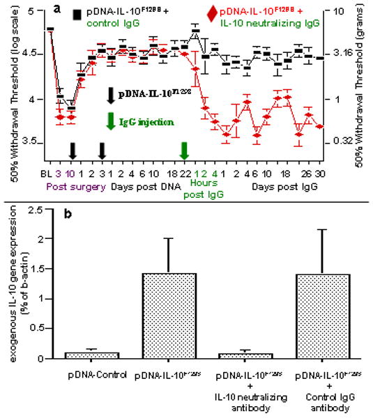
(a) Long term gene therapy stability requires IL-10 signaling. Long term therapeutic efficacy requires continuous IL-10 protein signaling following the second pDNA-IL-10F129S injection. Here, baseline (BL) responsivity to calibrated low threshold mechanical stimuli (von Frey test) was recorded prior to chronic constriction injury of the left sciatic nerve. Von Frey testing re-occurred through 62 days after surgery at time points indicated on the X-axis. Ten days after surgery, a time by which robust mechanical allodynia had developed, rats received 100 ug pDNA-IL-10F129S followed 3 days later by 25 ug pDNA-IL-10F129S (at time points indicated by two serial black Arrows on X-axis). A single injection (at green Arrow on X-axis) of IL-10 neutralizing antibody (0.2 ug; red diamonds; N=6, across 2 replications) led to the rapid and sustained re-expression of pain behavior as compared to pre-injection withdrawal thresholds (p<0.01 beginning at 2 hr following neutralizing antibody and lasting the remainder of the experiment). Control IgG injection (at green Arrow on X-axis) did not disrupt long term therapeutic efficacy (black squares; N=6, across 2 replications). Error bars indicate SEM. (b) Plasmid derived gene expression requires continuous IL-10 signaling. Rats were prepared as in Panel a. At 22 days following the second pDNA-IL-10F129S injection (100 ug followed by 25 ug), significant plasmid derived IL-10 expression is observed in cells contained in the lumbosacral CSF (second bar), compared to rats receiving 2 successive intrathecal injections (100 ug followed by 25 ug) of non-coding pDNA-Control (first bar) (p<0.05). The remaining rightward 2 bars represent data of separate groups of rats that also had received 2 successive intrathecal injections (100 ug followed by 25 ug) pDNA-IL-10F129S prior to challenge by intrathecal antibody. A single injection of IL-10 neutralizing antibody (0.2 ug; N=6, across 2 replications) given on day 22 post second pDNA-IL-10F129S injection (third bar) reduced plasmid derived IL-10 gene expression in lumbosacral CSF to background levels (equivalent to levels following pDNA-Control injections) within 4 days (the timepoint of CSF analysis shown here). Injection of an equivalent amount of control IgG antibody (N=6, across 2 replications) given under the same conditions has no effect on exogenous IL-10 gene expression in CSF as compared to the levels observed at day 22 post second pDNA-IL-10F129S injection (rightmost bar).