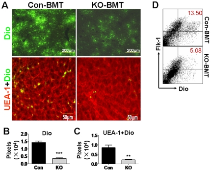
Figure 2. Lowered recruitment of the RBP-J-deleted EPC into the regenerating liver after PHx.
Normal mice were subjected to PHx. On the next day of the operation, mice were irradiated, and were transfused with Dio-labeled BM cells from the RBP-J deficient (KO-BMT) or the control (Con-BMT) mice. The recipient mice were analyzed 2 days after the BM transplantation (3 days after PHx). (A) The livers of the recipient mice suffering PHx were sectioned, stained for UEA-1, and were examined for Dio+ cells (upper) and UEA-1+Dio+ cells (lower). (B, C) Dio+ cells and UEA-1+Dio+ cells in (A) were quantitatively represented by corresponding pixels (bars = mean±SD; n = 4; ** P<0.01; *** P<0.001). (D) Livers were perfused with PBS. SECs were purified from the livers of the recipient mice using a kit, and were analyzed for Flk-1+Dio+ cells. The cytograph represented 4 independent experiments with the same results.

