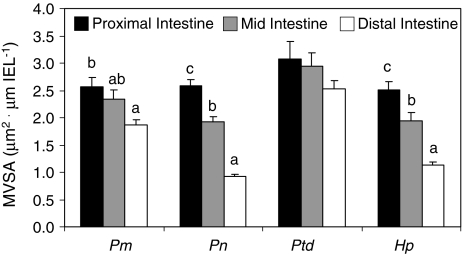
Fig. 4.
Microvilli surface area (MVSA, μm2) per length of intestinal epithelium (IEL, μm) in Panaque cf. nigrolineatus “Marañon” (Pm), P. nocturnus (Pn), Pterygoplichthys disjunctivus (Ptd), and Hypostomus pyrineusi (Hp). Values are means and error bars represent SEM. Intraspecific comparisons of MVSA among gut regions were made with ANOVA followed by a Tukey’s HSD with a family error rate of P = 0.05. Regional MVSA for a particular species that share a letter are not significantly different. No letters above gut regions (e.g., Pt. disjunctivus) indicate that there are no differences in MVSA among the intestinal regions for that species. Interspecific comparisons among species were not made

