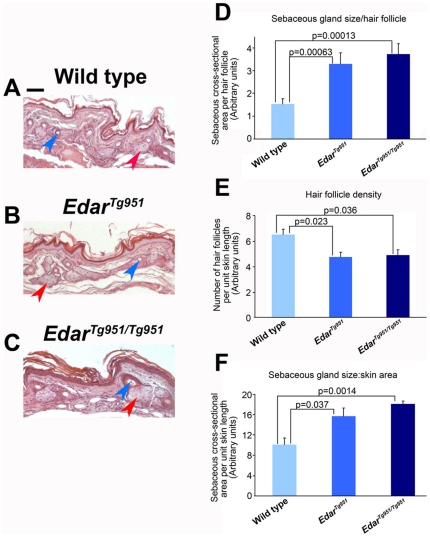
Figure 1. Increased sebaceous gland size in transgenic mice with elevated Edar signalling.
(A–C) Haematoxylin & eosin stained sections of the dorsal region of the hindfeet. A subset of sebaceous glands is indicated by red arrowheads and hair canals by blue arrowheads. (D) Quantification of cross-sectional area of sebaceous glands normalised to hair follicle number. (E) Hair follicle density in wild type and transgenic skin. (F) Quantification of cross-sectional area of sebaceous glands normalised to skin area. Scale bar indicates 50 µm.

