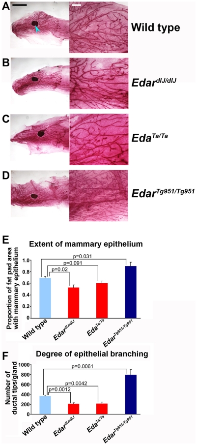
Figure 6. Mammary gland branching and epithelial growth extent is stimulated by Edar signalling.
Whole mount stained mammary glands of 6 week old (A) wild type, (B) EdardlJ/dlJ loss of function mutant, (C) EdaTa/Ta loss of function mutant and (D) EdarTg951/Tg951 gain of function transgenic. The lymph node within the fat pad is indicated by a blue arrowhead in (A). (E) Quantification of the extent of mammary epithelial infiltration into the fat pad. (F) Determination of epithelial branching, expressed as the total number of ductal termini per mammary gland for each genotype. Comparison of mutant to transgenic mammary gland morphometric measures gave p-values <0.01. For A-D the black scale bar on the left panels indicates 5 mm and the white scale bar on the right panels indicates 1 mm.

