Figure 1.

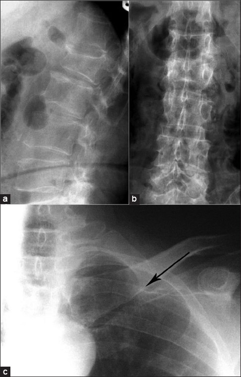
X-ray lumbosacral spine lateral view (a) and anteroposterior view (b) showing compression fracture of L2 vertebrae. X-ray of (L) shoulder with clavicle shows a thin hair line fracture at the medial end of left clavicle (solid arrow)

X-ray lumbosacral spine lateral view (a) and anteroposterior view (b) showing compression fracture of L2 vertebrae. X-ray of (L) shoulder with clavicle shows a thin hair line fracture at the medial end of left clavicle (solid arrow)