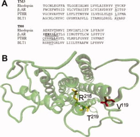
Figure 1.

Location of the mutated residues. A: Alignment of the TM3 and TM6 segments of rhodopsin, β2-adrenergic, PTH and BLT1 receptors. The sequences of human BLT1, bovine rhodopsin, human β2-adrenergic receptor, and opossum PTHR were aligned using Clustal W.24. The residues mutated in each receptor are underlined. The residues in the TM6 segment of the β2-adrenergic and PTH receptors directly involved in cation coordination are given in bold.19 B: Model of the 7TM helices showing the position of the mutated residues in TM3 and TM6. The model is presented from the cytoplasmic face. The residues in TM3 (V119) and TM6 (R218 and T219) that have been replaced by histidines are indicated. The figure in (B) was prepared in VMD.25
