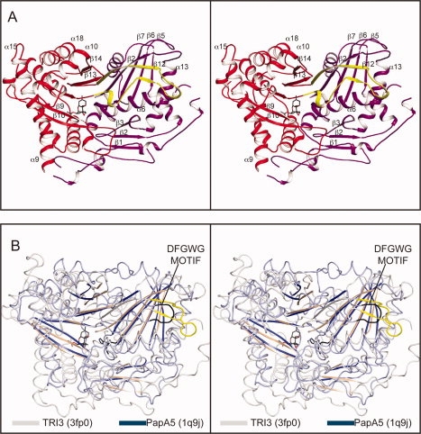
Figure 2.

Structural representations of TRI3. (A) Stereoview of TRI3 complexed with 15-decalonectrin (PDB accession number 3fp0). The N- and C-terminal domains are colored magenta and red, respectively, and the domain swapped β-strand 12 is colored yellow. Bound ligand 15-decalonectrin is colored dark gray. (B) Stereo overlay of TRI3 (colored white) and vinorine synthase, PDB ID 2bgh, (colored light blue). The loop between residues Asp362 and Gly366 of vinorine synthase are colored black and the corresponding loop residues, Glu449-Ser461, of TRI3 are colored yellow.
