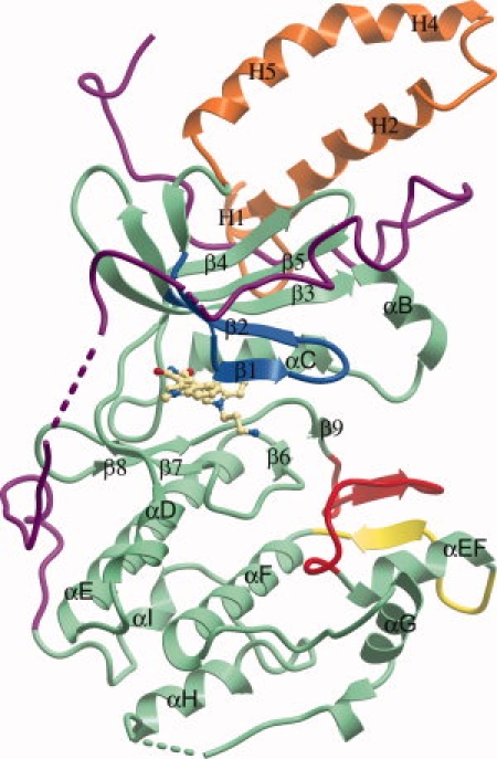
Figure 1.

Domain arrangement of DMPK. The BIM-8 inhibitor in the active site is shown as a ball-and-stick representation. The N-terminal kinase lobe is mostly above the BIM-8 inhibitor and the C-terminal kinase lobe mostly below. The C-terminal section of the protein is colored purple, the activation loop is colored red, the αEF/αF loop is colored yellow, and the glycine rich loop colored blue. The N-terminal helices involved in the dimerisation interface are colored orange.
