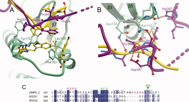
Figure 2.

Comparison of the binding of the C-termini of DMPK and ROCK1 to the N-terminal lobe. (A) The PXXP motif, the ROCK1 C-terminus is in yellow, and DMPK is colored as in Figure 1. (B) The turn motif. (C) Sequence alignment of DMPK and the Rho kinases over the region covering the PXXP, active-site tether and turn motifs. The VSGGG insertion present in isoforms 1 and 2 of DMPK is indicated with red lettering.
