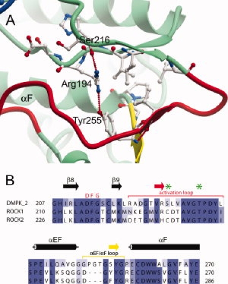
Figure 5.

(A) Activation segment of DMPK, coloured as in Figure 1. (B) Partial sequence alignment of the activation loop and αEF/αF loop regions of DMPK against the Rho kinases 1 and 2, which are colored as in (A). The residues equivalent to those phosphorylated in MRCK are indicated with green stars. A full sequence alignment of the DMPK subfamily can be seen as Supplementary Figure 1.
