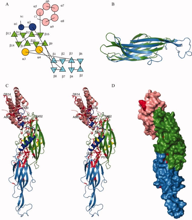
Figure 2.

Structure of pT26-2-6: (a) Topology diagram of pT26-6p. The repeated β-sandwich domains are colored blue and green and the helical bundle pink. The N-terminal helical peptide is in deep blue and the helical insertion in the fist sandwich domain in yellow. (b) Superposition of the two β-sandwich domains. (c) Cartoon stereo view representation using the same color scheme as (a) conserved residues are colored in red, some of them are labeled. (d) Surface representation of conserved residues (in red).
