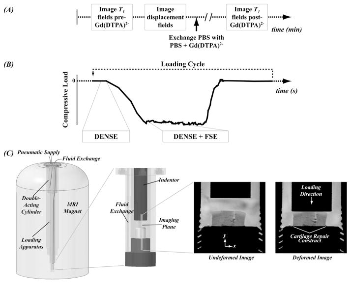
Figure 2.
Functional MRI analyses determined deformation and [GAG] fields in the tissue and required precise timing methods. Timing of fluid exchange actions, MRI T1 measurements, and mechanical loading allowed for computation of deformation fields and [GAG] (A). Integration of DENSE and FSE pulse sequences during cyclic mechanical loading allowed for measurement of deformation fields (B). A custom MRI-compatible bioreactor was used for application of mechanical loading and fluid exchange (C). Standard MRI (FSE alone) generated undeformed and deformed images with similar proton density in the construct and surrounding tissue and did not allow for functional assessment of the tissue-engineered construct.

