Figure 1.
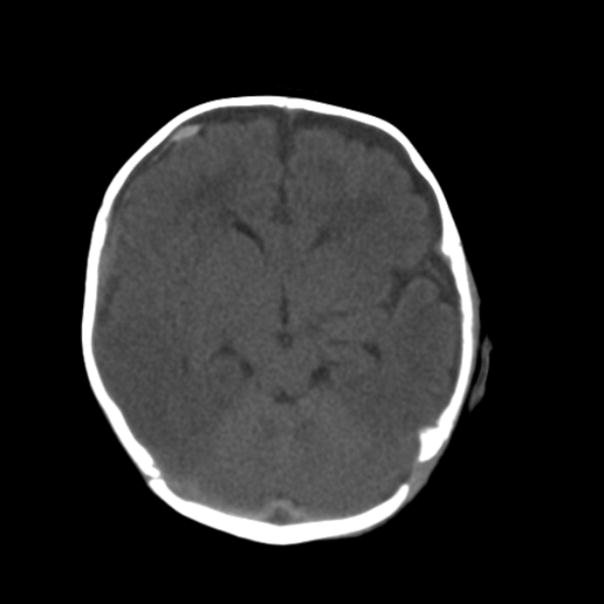
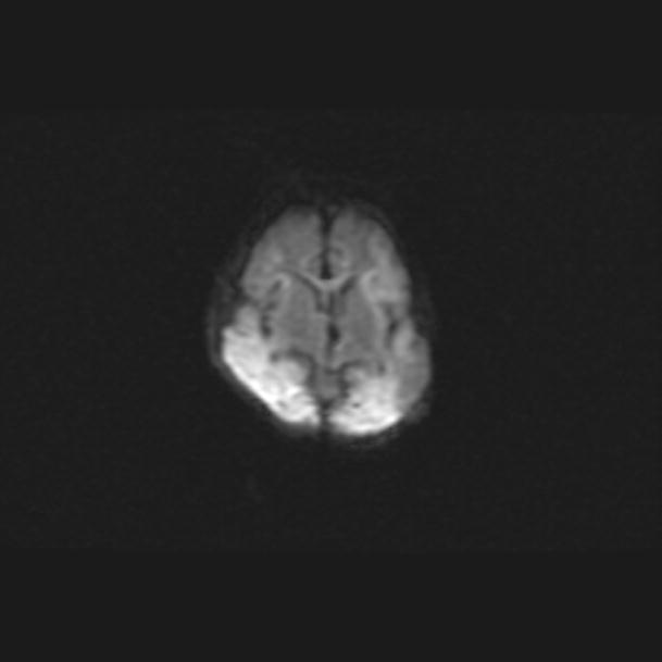
Figure 1A. Noncontrast head CT in a child with high clinical suspicion of child abuse shows right frontal subdural hematoma but no evidence of ischemia. Figure 1B. Diffusion weighted MR sequence (ADC maps showed corresponding low signal) in the same patient demonstrates ischemia in bilateral parietal lobes not detected on the initial head CT.


