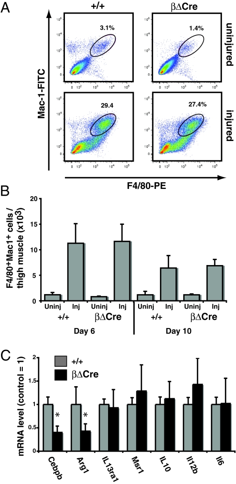
Fig. 6.
Infiltrating macrophage phenotype in injured βΔCre muscle. (A) Phenotypic analysis from +/+ and βΔCre muscle uninjured and injured 6 days after CTX injection by flow cytometry for the presence of Mac-1+F4/80+ cells. Plots are representative of four independent experiments and numbers represent the frequency of cells in the indicated gates. (B) Total mononuclear cells were isolated from injured and control thigh muscle and analyzed for expression of F4/80 and Mac-1 by FACS. The total number of recovered F4/80+Mac-1+ cells is indicated for each condition (n = 2 for uninjured samples; N> = 5 for injured samples). Error bars indicate standard deviations. (C) F4/80+Mac-1+ cells were sorted from day 6 injured muscle, and gene expression analyzed by real-time PCR (N> = 5/genotype). Data are presented as the mean ± SD normalized to the +/+ value (= 1). Asterisks (*) indicate P < 0.05 (Student's t-test).

