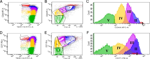
Fig. 3.
Flow cytometric analysis of bone marrow cells. (A–C) Bone marrow cells labeled with antibodies against TER119 and CD44. (A) plot of CD44 versus TER119. (B) Plot of CD44 versus FSC of all TER positive cells. Note that 5 distinct clusters can be distinguished. (C) The CD44 expression levels in the gated cell population. Note the progressive decrease of CD44 surface expression from region I to region V. (D–F) Bone marrow cells labeled with antibodies against TER119 and CD71. Note that cells from regions I to III express similar levels of CD71.

