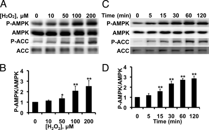
Fig. 1.
H2O2−mediated AMPK phosphorylation in endothelial cells. Shown in this figure are the results of immunoblots analyzed in endothelial cells treated with H2O2. (A) Representative immunoblot from a dose-response experiment analyzed in cells stimulated with the indicated concentrations of H2O2 for 30 min and probed with antibodies as shown; (B) pooled data from five independent experiments, analyzing the intensities corresponding to phospho-AMPK and total AMPK by quantitative chemiluminescence. (C) Representative time course experiment in BAECs treated with 200 μM H2O2 for the indicated times and analyzed in immunoblots probed with antibodies as shown; (D) pooled data from five independent experiments. *, P < 0.05, and **, P < 0.01 by ANOVA.

