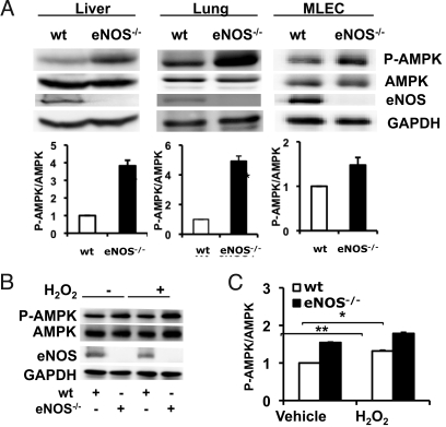
Fig. 6.
AMPK phosphorylation in tissues and cells from eNOS−/− mice. This figure shows immunoblot analyses of liver, lung, or endothelial cells from wild-type (wt) or eNOS−/− mice. (A) Liver, lung tissues and isolated lung endothelial cells (MLECs) from wild-type and eNOS−/− mice were analyzed in immunoblots probed with antibodies as indicated. Shown below are pooled data from at least three experiments quantitating AMPK phosphorylation; ***, P < 0.001. (B) MLECs from wild-type or eNOS−/− mice were treated with H2O2 (100 μM for 30 min). The experiment shown is a representative of five similar experiments showing that MLECs from eNOS−/− mice have increased basal as well as H2O2-stimulated AMPK phosphorylation relative to MLECs from wild-type mice; pooled data are also shown,*, P < 0.05.

