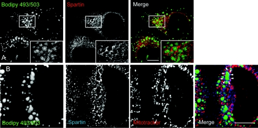
Figure 3. Endogenous spartin can be recruited to lipid droplets.
(A) HeLa cells were treated with oleic acid (200 μM for 24 h) and lipid droplets were labelled by incubation with BODIPY 493/503. Cells were then fixed and labelled for spartin. A spartin signal often surrounded the lipid droplets and, where lipid droplets clustered, a honeycomb appearance was seen (see boxed area; inset shows a magnified image of the boxed area). (B) A cell in which lipid droplet formation was induced in the same way as in (A). The cell was labelled with the mitochondrial marker MitoTracker, as well as with α-spartin and BODIPY 493/503. The spartin signal surrounds many of the lipid droplets; an example is indicated by the arrowheads. The spartin label around the lipid droplets shows minimal co-localization with MitoTracker. Scale bars=10 μm.

