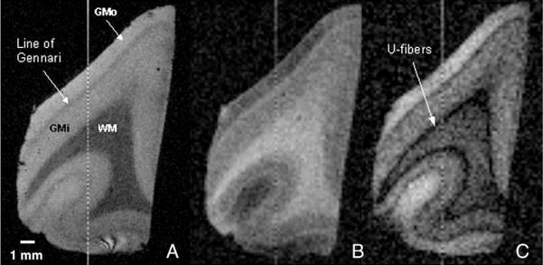
Fig. 4.
MR images with T2-weighting (A), T1-weighting (B), and intermediate weighting at 14 T of an LFB-stained, LiCO-differentiated sample of human visual cortex. Three regions of interest used for analysis are clearly visible in all images: white matter (WM), inner gray matter (GMi), comprised of layers IVc–VI, and outer gray matter (GMo), comprised of layers I–IVa. The Line of Gennari, layer IVb that is heavily myelinated in visual cortex, is also prominently displayed. In image C, the subarcuate fibers, or “u-fibers,” are also resolvable and appear darker than the underlying white matter. Images have 150-micron isotropic resolution and were acquired using (A) a gradient echo sequence (TR/TE=80/15.2 ms, Θ=30°); (B) an inversion-prepared spin echo sequence (TR/TE/TI=5000/5.96/320 ms); and (C) an inversion-prepared spin echo sequence (TR/TE/TI=5000/5.96/120 ms).

