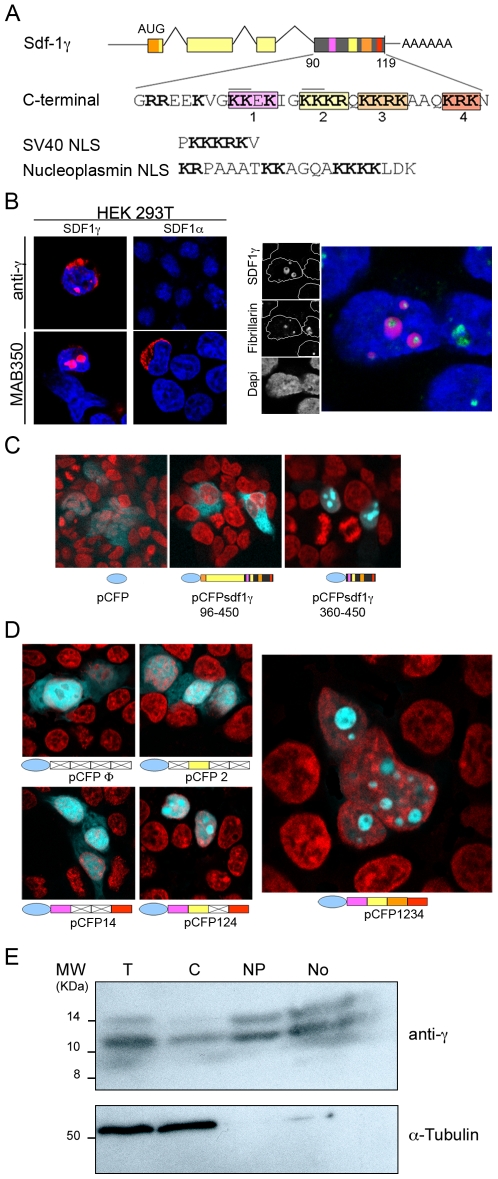
Figure 2. The Sdf-1γ carboxy-terminal region contains a nucleolar localization signal.
(A) Schematic of the Sdf-1 locus showing the predicted Sdf-1γ exon structure, based on the annotated Sdf-1γ mRNA sequence from brain, and detail of the unique Sdf-1γ C-terminal exon 4. Basic amino-acid residues (Arg/Lys) are shown bold, and clusters of basic residues are boxed and numbered. SV40 and nucleoplasmin NLS sequences are shown for comparison. (B) Left panel. Confocal immunofluorescence of HEK293T cells transfected with plasmids pSdf1γ–30–450 (Sdf-1γ) and pSdf1α–30–450 (Sdf-1α). Subcellular localization of overexpressed Sdf-1 proteins (red) was detected with pan anti-Sdf-1 (MAB350) or specific anti-Sdf-1γ (anti-γ). Right panel. Co-labeling of the nucleolar protein fibrillarin and pSdf-1γ by confocal immunofluorescence. HEK293T cells were transfected with plasmid pSdf1γ–30–450 and immunostained for fibrillarin and Sdf-1γ. Individual and merged images of a representative field are shown on the left, and the inset regions are shown at high magnification on the right to show the localization of Sdf-1γ (green) and fibrillarin (red) in granular and fibrilar nucleololar regions, respectively. In both panels nuclei are stained with DAPI (blue). (C) Fluorescence images of HEK293T cells transfected with the indicated plasmids encoding cerulean fluorescent protein (CFP) fused to full-length Sdf-1γ (left) or the Sdf-1γ specific carboxy-terminus (center); The right panel shows results with unfused CFP. CFP fluorescence is shown blue, and nuclei are stained with TOPRO-3 (red). (D) Fluorescence images of HEK293T cells transfected with CFP fusions of wild-type or mutated versions of the Sdf-1γ C-terminal domain. Unmutated clusters of basic residues are colored as in A, and mutation (Lys/Arg→Ala) of the clusters is shown by the white/crossed boxes. Fluorescence signals are as in (C). Arrowheads indicate nucleoli. (E) Western blot of subcellular fractions of cell extracts. Cell equivalents were loaded on each lane. T, whole cell extract; C, cytoplasm; Np, nucleoplasm; No, nucleoli. Sdf-1γ was stained with anti-γ and developed with HRP-goat anti rabbit secondary antibody.

