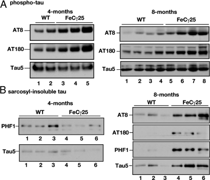
Fig. 1.
Increased phosphorylation and age-dependent aggregation of tau in FeCγ25 mice. (A) Left panel: Hippocampal brain lysates from 4-month-old wild-type mice (lanes 1, 2) or FeCγ25 transgenic mice (lanes 3–5) were separated by SDS/PAGE and Western blotted using AT-8 (top row), AT-180 (middle row), or Tau5 (lower row) to detect phospho-tau and total tau, respectively. Note the elevated levels of phospho-tau in FeCγ25 mice compared to wild-type controls. In contrast, total tau levels were unaltered. Right panel: Hippocampal brain lysates from 8-month-old wild-type mice (lanes 1–4) or FeCγ25 transgenic mice (lanes 5–8) were separated by SDS/PAGE and Western blotted using AT-8 or AT-180 antibodies as described above. The phospho-tau levels remained elevated at 8 months of age in FeCγ25 mice while the total tau levels remained essentially unchanged. (B) Left panel: Lysates from brain cortices from wild-type (lanes 1–3) or FeCγ25 (lanes 4–6) mice were subjected to a sarcosyl extraction protocol and the detergent-insoluble fractions were Western blotted using PHF1 or Tau5 antibodies. There were no consistent differences in the amounts of detergent-insoluble tau at 4 months of age between FeCγ25 and control mice. Right panel: Cortical brain lysates from 8-month-old wild-type (lanes 1–3) or FeCγ25 (lanes 4–6) mice were subjected to a sarcosyl extraction protocol and the detergent insoluble fractions were Western blotted using indicated antibodies. Note the increased amounts of both phospho-tau and total tau in the sarcosyl-insoluble fraction of FeCγ25 mice. Each lane represents an individual mouse.

