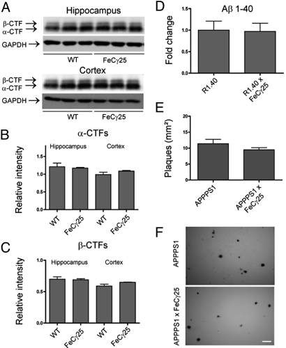
Fig. 6.
AICD does not alter APP processing or increase Aβ levels. (A) Western blots from 3–4-month-old hippocampal and cortical lysates for APP carboxy-terminal fragments with APP anti-carboxy terminal 0443 antibody showing representative alpha and beta-cleaved bands and GAPDH loading controls. Each lane represents an individual mouse. (B) α-CTFs were quantified by normalizing the band intensity to that of GAPDH to determine the relative intensity for both hippocampal and cortical samples, with no significant differences in protein levels. (C) β-CTFs were quantified in the same manner as described for α-CTFs and show no significant differences in relative protein levels. (D) ELISA to detect Aβ 1–40 from 3–4-month-old hemi brains of R1.40 or R1.40 × FeCγ25 demonstrated no significant differences between groups. (E) 6E10-positive plaques were counted from 3–4-month-old APPPS1 or APPPS1 × FeCγ25 mice, with no apparent differences in the number of plaques. (F) Representative plaque deposition in APPPS1 and APPPS1 × FeCγ25 mice from cortices. All data expressed as mean ± SEM, n = 3 for all groups. [Scale bar in (F), 100 μm.]

