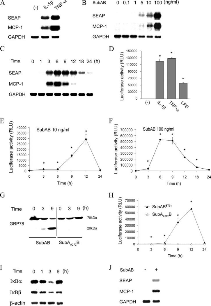
FIGURE 2.
Transient activation of NF-κB and induction of NF-κB-dependent gene expression by SubAB. A, NRK-52E cells were stably transfected with pNFκB-SEAP, and NRK/NFκB-SEAP reporter cells were established. The cells were stimulated with 1 ng/ml IL-1β or 10 ng/ml TNF-α and subjected to Northern blot analysis of SEAP and MCP-1. Expression of GAPDH is shown at the bottom as a loading control. B and C, NRK/NFκB-SEAP cells were stimulated with serial dilutions of SubAB for 6 h (B) or 10 ng/ml SubAB for indicated time periods (C) and subjected to Northern blot analysis. D, NRK-52E cells were stably transfected with pNFκB-Luc, and NRK/NFκB-Luc cells were established. The cells were stimulated with IL-1β, TNF-α, or 1 μg/ml LPS and subjected to chemiluminescent assay to evaluate luciferase activity. RLU, relative light unit. E and F, NRK/NFκB-Luc cells were treated with 10 ng/ml (E) or 100 ng/ml (F) of SubAB for up to 24 h and subjected to luciferase assay. G, Cells were treated with SubAB or its inactive mutant SubAA272B for up to 9 h and subjected to Western blot analysis of GRP78. H, NRK/NFκB-Luc cells were stimulated with endotoxin-free SubABET– (10 ng/ml) or SubAA272B (10 ng/ml) for up to 24 h and subjected to chemiluminescent assay. I, NRK-52E cells were treated with 100 ng/ml SubAB for up to 6 h and subjected to Western blot analysis of IκBα and IκBβ. J, SM/NFκB-SEAP cells were treated with 100 ng/ml SubAB for 9 h and subjected to Northern blot analysis of SEAP and MCP-1. In D–F and H, assays were performed in quadruplicate, and data are expressed as means ± SE. Asterisks indicate statistically significant differences (p < 0.05).

