
Figure 4. Expression and angiogenesis inhibition by the human endostatin-angiostatin fusion protein in Suit-2 cells infected with VVhEA in vitro.
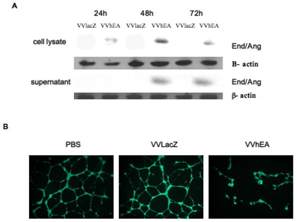

A, Endostatin-angiostatin fusion protein expression in Suit-2 cells. Suit-2 cells were infected with 1 PFU/cell of VVhEA or VVlacZ. Cells and supernatant were harvested 24, 48 and 72 hours later. Western blots were performed as described in Materials and Methods. All fusion protein bands were between 70 and 80 kDa and β actin around 45 kDa. Although intracellular transgene expression was found in the cells by 24 hours following infection, endostatin-angiostatin fusion protein was detected in the supernatant only after 48 hours; B and C, Inhibition of HUVEC tube formation. HUVEC were seeded in Matrigel with 10μg of supernatant harvested and concentrated 72 hours after Suit-2 cells were infected with 1 PFU/cell of VVhEA or VVlacZ. 16 hours later, 400 μg Calcein AM was added and fluorescence microscopy used to assess the number of fully formed HUVEC tubes, which were compared to mock-infected controls and each other by one-way ANOVA; D, Inhibition of HUVEC proliferation, Mean cell survival ± SEM as a percentage of uninfected HUVEC by MTS assay 96 hours post-treatment with 10μg/ml of supernatant harvested from Suit-2 cells infected with 1 PFU/cell of VVhEA or VVlacZ for 72 hours. Mean results were compared by one-way ANOVA with Bonferonni post-hoc testing.