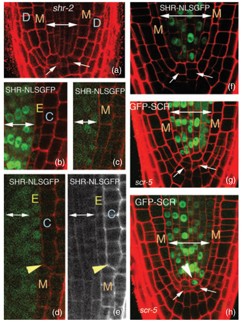
Figure 2. Cell-autonomous SHR does not rescue shr-2.
(a) Longitudinal confocal section through the tip of a shr-2 root. Arrows indicate abnormal QC morphology.
(b) Longitudinal confocal section through the tip of a wild-type root expressing SHRpro–SHR–NLSGFP.
(c–e) Longitudinal confocal micrographs of shr-2 plants expressing SHRpro–SHR–NLSGFP. (c) The cell-autonomous SHR–NLSGFP protein is unable to rescue either the radial patterning or QC morphology. However, in (d), rescue of radial patterning is coincident with movement of the SHR–NLSGFP protein into the ground tissue. The yellow arrowheads in (d) and (e) indicate the wall between the endodermis and cortex. Notice that above the yellow arrowhead there are two distinct ground-tissue layers, and SHR–NLSGFP is clearly detected in the endodermal layer. (e) The red-channel image of the root in (d) is shown in gray scale to allow easier visualization of the rescue of the endodermal layer.
(f) shr-2 root expressing SHRpro–SHR–NLSGFP. This root has aberrant radial patterning and QC morphology, but normal root length despite a lack of detectable GFP in the QC cells.
(g,h) scr-5 roots expressing SHRpro–GFP–SCR. Expression of SCR in the stele does not rescue the scr-5 radial patterning defects, but these roots do have normal lengths. In (h), there is an indication of divisions at the base of the stele (arrowhead) above the abnormal QC (arrows) forming a population of cells that morphologically resemble a new QC.
Double-headed arrows delineate stele cells throughout. M, mutant ground-tissue layer; D, epidermis; E, endodermis; C, cortex.

