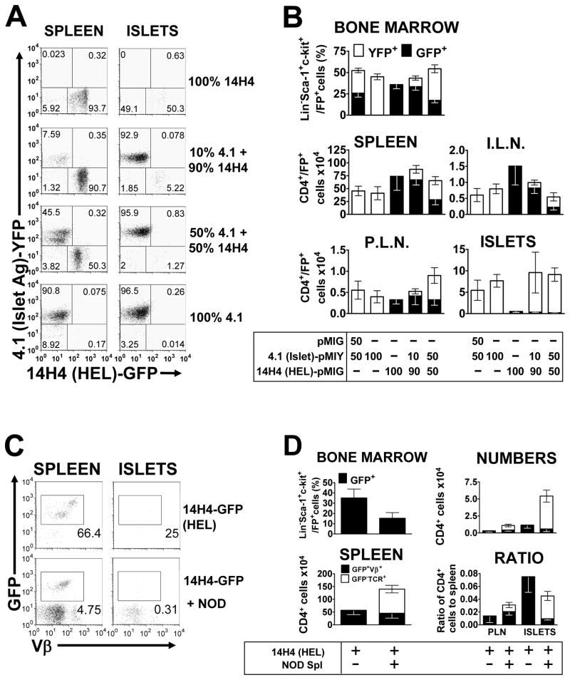
Figure 2. HEL-specific “Bystander” CD4+ T cells do not accumulate in the pancreas.
Double retrogenic mice, with a diabetogenic T cell labeled with YFP and a HEL-specific “bystander” T cell labeled with GFP. Mice were analyzed 4.5–6.5 weeks post transfer, with the bone marrow, spleen, inguinal lymph nodes, pancreatic lymph nodes and pancreatic islets being extracted, processed, cells counted and subjected to flow cytometric analysis. (A) Representative flow cytometric dot plots from B, gated on live and CD4+ T cells. (B) Mice received bone marrow transduced with either 4.1 or 14H4, or both, at a 50:50 ratio, or a 10:90 ratio. One group received 4.1-YFP bone marrow and bone marrow transduced with empty pMIG vector as a control. Top panel; mean (±SEM) percentage of stem cells for each T cell in the bone marrow, as measured by percentage GFP/YFP expression on Lineage−Sca-1+c-kit+ cells (n=10). Middle and bottom panels, mean (±SEM) numbers of each T cell in the spleen (n=9–10), inguinal lymph nodes (I.L.N, n=7–9), pancreatic lymph nodes (P.L.N., n=6–9) and pancreatic islets (ISLETS, n=10), respectively, as calculated from cell counts and the percentage CD4+FP+ live cells. (C) Representative flow cytometric dot plots from D, showing analysis of T cells from 14H4 HEL-specific mice, injected with 10×106 whole splenocytes from 13 week-old NOD mice, two weeks after bone marrow engraftment, and analyzed 4.5–6.5 weeks post-transplant. Plots are gated on live cells and CD4+ cells. (D) Left panels; percentage of stem cells for each T cell in the bone marrow (n=7–10), as measured by percentage GFP expression on Lineage−Sca-1+c-kit+ cells and total numbers of T cells in the spleen. Right panels show the numbers and ratio to the numbers in the spleen for the pancreatic islets (ISLETS) and pancreatic lymph nodes (P.L.N.), respectively (n=5–9). All figures show mean (±SEM) and numbers were calculated from cell counts and the percentage CD4+GFP+Vβ+ live cells for the retrogenic T cells and the percentage CD4+GFP−TCR+ live cells for the NOD T cells.

