
Figure 2. EMs of kDNA networks.
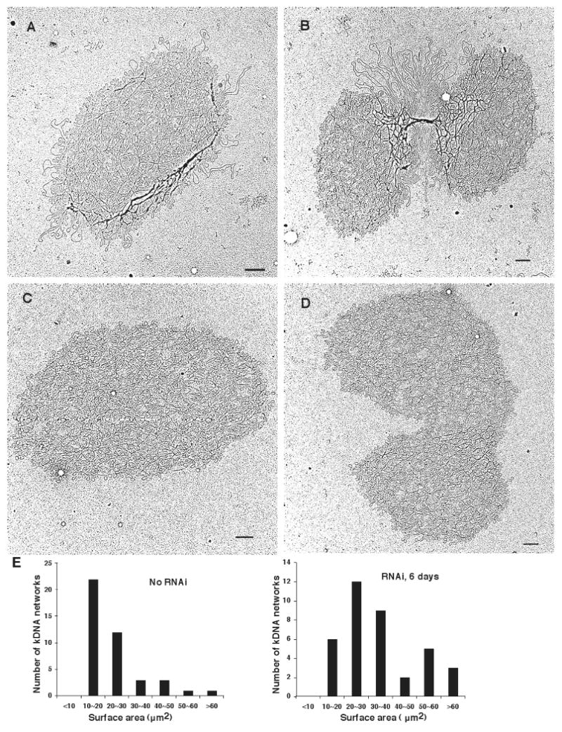
(A) and (B), kDNA isolated from wild-type cells. (C) and (D), kDNA isolated from TbPIF2 RNAi cells (4 days after induction). Networks in B and D are double-size and are undergoing segregation. Bar, 500 nm. (E) Bar graphs showing surface areas of randomly-chosen networks (including unit-size and double-size) from uninduced cells (left panel) and RNAi cells (6 days, right panel). Maxicircle loops on network periphery were not included in surface area measurement.