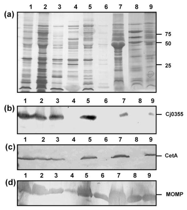
Fig. 1.
Outer-membrane fractions of C. jejuni strain 81–176 from nine purification methods. (a) Silver-stained SDS-PAGE analysis of OMP preparations (5 μg). Lanes: 1, glycine extraction; 2, differential detergent extraction using Triton X-100; 3; serial extraction using 0.1 M Tris/HCl, pH 7.8; 4, spheroplasts isolated using lysozyme treatment; 5, spheroplasts isolated using sonication and lysozyme treatment; 6, carbonate washed spheroplasts; 7, carbonate extraction; 8, Sarkosyl extraction; 9, sucrose gradient fraction 14. Molecular masses are indicated on the right (in kDa). (b–d) Immunoblots of the outer-membrane preparations using the cytoplasmic marker anti-Cj0355 (b), the inner-membrane marker anti-CetA (c) and the outer-membrane marker anti-MOMP (d). All antibodies were used at 1: 1000 dilution. Loading for panels (b)–(d) is the same as for panel (a).

