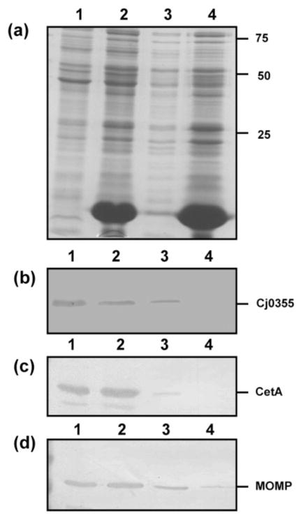
Fig. 3.
Analysis of the preparative steps for spheroplasts of C. jejuni strain 81–176 using the lysozyme method. (a) Silver-stained SDS-PAGE gel of the preparative steps. Lanes: 1, whole-cell lysate; 2, post-lysozyme treatment supernatant; 3, inner membrane isolated from spheroplasts; 4, supernatant following centrifugation to remove unlysed cells (contains mostly outer membrane). Ten microlitres of each of the preparative steps was loaded per lane. Molecular masses are indicated on the right (in kDa). (b–d) Immunoblots of the lysozyme spheroplast preparatory samples using the cytoplasmic marker anti-Cj0355 (b), the inner-membrane marker anti-CetA (c) and the outer-membrane marker anti-MOMP (d). All antibodies were used at 1: 1000 dilution. Loading for panels (b)–(d) is the same as for panel (a).

