
Figure 1. Assessment of myocardial function after cell transplantation by echocardiography.
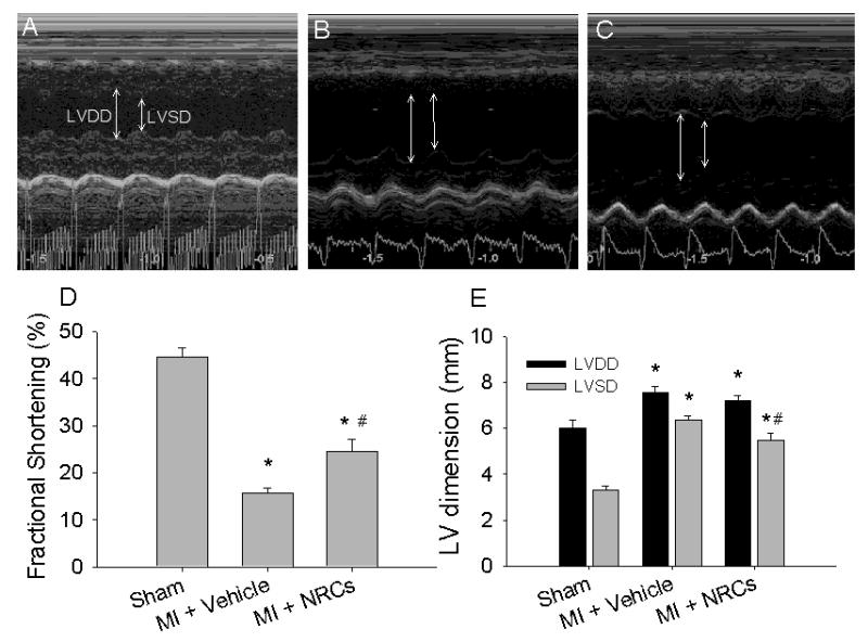
Representative left-ventricular M-mode images from control animals (A, sham, n = 6), and infarcted animals that received either vehicle (B, non-treated infarcted, n = 9) or cells (C, grafted, n =10). Arrows indicate chamber dimension at end-diastole (LVDD) and end-systole (LVSD). Fractional shortening (D) was greatly reduced with MI, but the functional decline was attenuated by cell transplantation. Both diastolic and systolic LV dimensions increased after MI (E). No difference was found in LV diastolic dimension (E, LVDD) between non-treated infarct or grafted hearts, but LV systolic dimension (E, LVSD) was reduced in grafted hearts. Values are means ± S.E.M.; * p < 0.05 versus sham; # p < 0.05 versus MI + vehicle.