
Figure 2. Changes in the number of dopaminergic neurons induced by progesterone action on ESC.
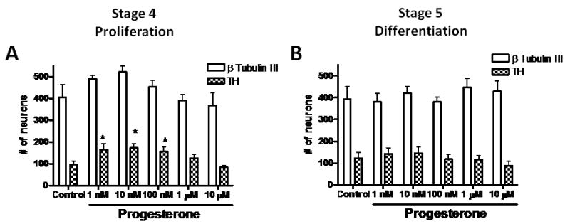
Quantification of the total number of β-Tubulin III and TH-positive cells from 8 fields from 3-6 independent experiments made in duplicate. Cells were incubated in progesterone-free N2 medium, with different progesterone concentrations during proliferation (A) or differentiation stages (B). Results are expressed as mean ± SEM. *P < 0.05 as compared with their corresponding control (cyclodextrin) group.