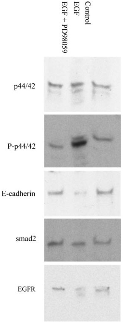
Figure 2. MAPK mediates the downstream affect of EGF in A549 cells.
Western blot analysis of the EGFR, MAPK (p44/42), phosphorylated MAPK (P-p44/42), E-cadherin, and smad-2, in lysates of A549 cells treated with EGF (200 ng/mL) or EGF and PD98059 (50 µM). Seventy micrograms of protein was loaded per well. In EGF treated A549 cells, the increase in phosphorylated MAPK signal is reversed by pretreatment with PD98059.

