
Figure 9.
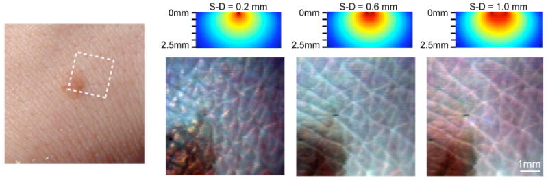
(online color at: www.lpr-journal.org) Laminar Optical Tomography of Skin. (left) Photograph of benign mole on the back of the hand. (right) Raw LOT reflectance images acquired using 488 nm, 532 nm, and 635 nm lasers. (above) simulations of sensitivity functions for each source-detector separation (S-D) (optical properties: μa = 0.7 mm−1 and at 532 nm). The narrowest source-detector separation accentuates superficial folds of the stratum corneum. Wider separations reveal folds in the epidermis, and sub-surface structures of the mole. Modified from [36].