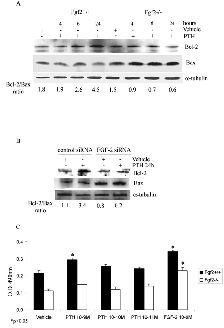
Fig 7. Effect of PTH on Bcl-2/Bax ratio in COBs from Fgf2+/+ and Fgf2−/− mice.
Expression levels of Bcl-2 and Bax in COBs were analyzed by Western blotting as described under methods. A, COBs from Fgf2+/+ and Fgf2−/− were treated with vehicle or PTH 10−9 M from 4 h to 24 h. Proteins (5 µg) from each sample were subjected to SDS–PAGE, transferred to PVDF membrane and probed with a polyclonal anti-Bcl-2 antibody or with a polyclonal anti-Bax antibody. Filters were stripped and reprobed with a monoclonal anti-α-tubulin to show equal amount of loading. B, Cells were transfected with FGF-2 siRNA or control siRNA as described above. After transfection, cells were serum deprived for 24h and treated with PTH 10−9 M from another 24h. Proteins (5 µg) from each sample were subjected to SDS–PAGE, transferred to PVDF membrane and probed with a polyclonal anti-Bcl-2 antibody or with a polyclonal anti-Bax antibody; filters were stripped and reprobed with a monoclonal anti-α-tubulin antibody to show equal amount of loading. C, MTS assay. Dose-response effect of PTH on the metabolic activity of viable COBs from Fgf2+/+ and Fgf2−/− mice. Cells were 24 h serum deprived and treated with PTH, from 10−9 to 10−11 M, or vehicle for 24 h. Other cells were treated with FGF-2 (10−9 M). Values are mean ± SEM for three different experiments. Note the significant increase of cell viability induced by PTH particularly at the concentration of 10−9 M only in Fgf2+/+ mice. *p < 0.05.

