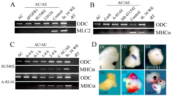
Figure 4. FGF and Nodal signaling are required during the first hour of cardiac induction.
(A) AC/AE conjugates were made with animal cap expressing ΔFGFR1 or with uninjected animal cap that were continuously incubated with SU5402 (50 µM) or U0126 (35 µM) from the time of conjugation, or were treated with DMSO (Control). MLC2 expression was analyzed at st. 34. (B) CerS (injected in animal cap) or continuous incubation with SB-431542 (75 µM) or A-83-01 (75 µM) block induction of MHCα. (C) FGF and Nodal signaling are only required the first hour of contact between AC and AE. (D) In situ hybridisation analysis of cardiac TnI expression in AC/AE conjugates. D1,2- control embryo and AC/AE explant. 83% (n = 23) explants are cTnI+. D3- expression of ΔFGFR1 in animal caps leads to a greatly reduced expression of cTnI in 89% (n = 19) explants. D4- 0/15 of CerS explants express cTnI. D5- mosaic expression of ΔActRI (red) in animal caps prevents expression of cardiac TnI (0/11 cTnI+ explants in ΔActRI-injected cells), but not in neighboring tissue with intact Nodal signaling (5/11 cTnI+ explants; arrow in D5). D6- constitutively active Alk4* receptor induces cardiac expression cell-autonomously in animal caps (4/4 cTnI+, n = 12).

