
FIG. 5.
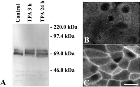
Effects of TPA on Cx56 and PKCγ in cultured chicken embryonic lens cells. (A) Immunoblot of Cx56 from homogenates of lentoid-containing cultures that were left untreated (Control) or that were treated with 200 nM TPA for 3 or 24 h. The migration positions of the molecular mass standards are indicated. (B, C). Photomicrographs show the distribution of anti-PKCγ immunoreactivity in lentoid cells left untreated (B) or treated with 200 nM TPA for 30 min (C). TPA treatment activated PKCγ (as demonstrated by its translocation to the plasma membrane) and produced a change in the immunoblot pattern of phosphorylated Cx56 forms. Bar, 12 μm in B and 15 μm in C.