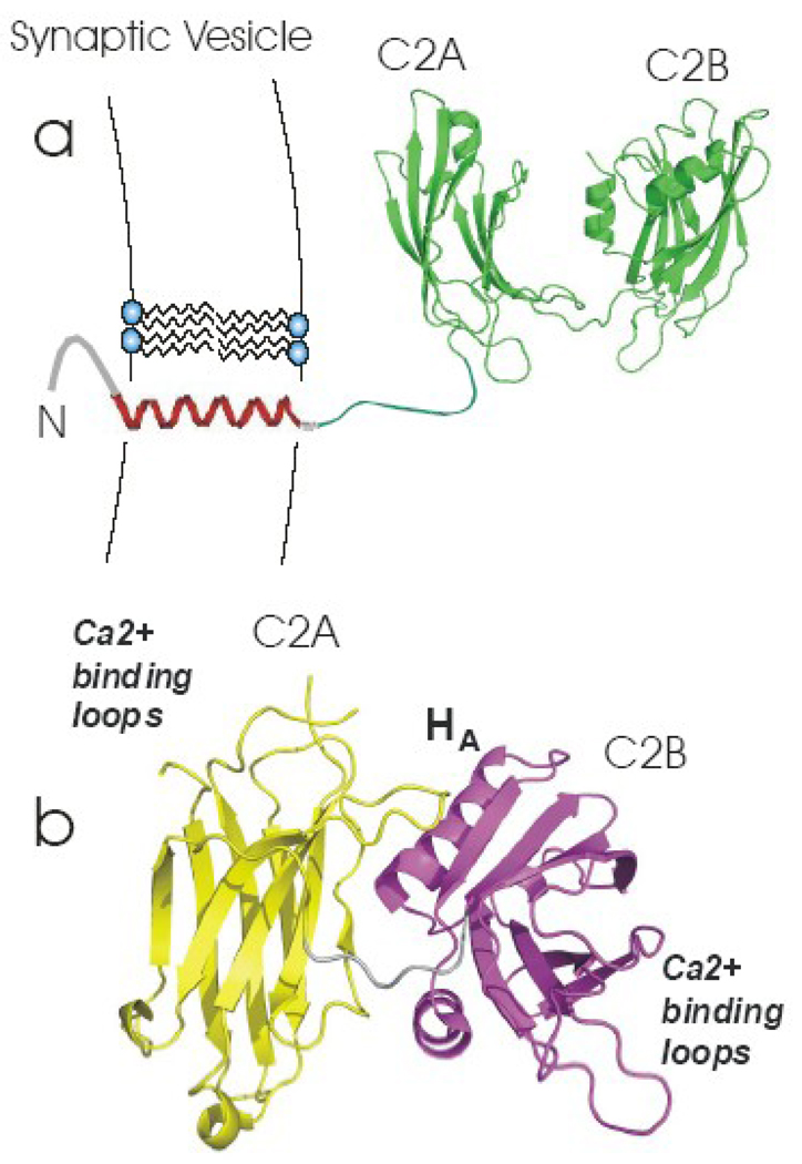
Figure 1. Models for synaptotagmin 1.
(a) Synaptotagmin 1 is a membrane protein containing two C2 domains, which is anchored to the synaptic vesicle membrane by a single transmembrane helix. This model was built from models for the isolated C2A(PDB:1BYN) and C2B(PDB:1K5W) domains, which were connected with an unstructured linker using InsightII. (b) Crystal structure of the soluble fragment of synaptotagmin 1 (PDB:2R83). The C2A and C2B domains are shown in yellow and magenta.

