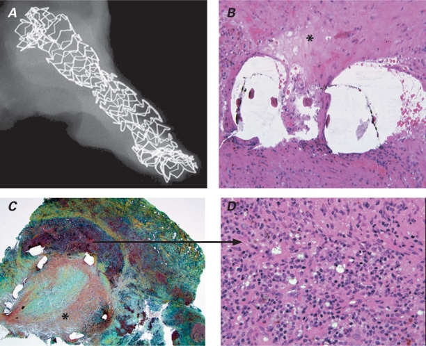
Fig. 5 A) Scanning electron microscopic view of the right coronary artery (RCA) stent. B) Hematoxylin & eosin stain (orig. ×40) of RCA pseudoaneurysm, in which the asterisk (*) shows stent struts with overlying thrombus. C) Photomicrograph (H & E, orig. ×10) of the RCA pseudoaneurysm, in which the asterisk (*) denotes organizing luminal thrombus, and D) the arrow demonstrates a magnified view (H & E, orig. ×60) of the arterial media with chronic inflammation. Findings are consistent with Behçet syndrome.

An official website of the United States government
Here's how you know
Official websites use .gov
A
.gov website belongs to an official
government organization in the United States.
Secure .gov websites use HTTPS
A lock (
) or https:// means you've safely
connected to the .gov website. Share sensitive
information only on official, secure websites.
