
Figure 6.
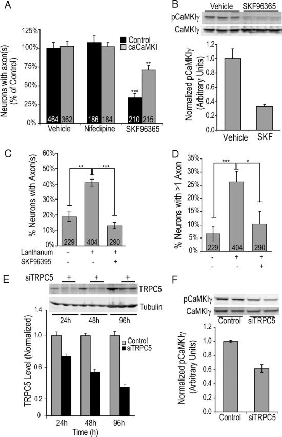
Regulation of CaMKIγ and axon formation by TRPC channel. A, Low-density hippocampal neurons electroporated before plating were incubated for 46 h (2 h after plating until fixation at 48 h) in culture with vehicle, the L-type channel blocker nifedipine (10 μm), or the TRPC blocker SKF96395 (3 μm). Neurons fixed at 48 h were stained with the axonal marker Tau-1. Quantification of results from three independent experiments is shown. B, Representative blot showing regulation of CaMKIγ by TRPC antagonist, SKF96365. P0 hippocampal neurons were electroporated with Flag-CaMKIγ before plating and 24 h later exposed to 50 μm SKF96365 for 60 min. Lysates were immunoblotted with anti-phospho-CaMKI antibody (top blot) or anti-Flag antibody to detect total CaMKIγ (bottom blot). The bottom graph shows average normalized phospho-CaMKIγ. C, D, Graphs depict quantification of axon formation (C) and induction of multiple axons (D) in E18 neurons that were incubated for 20 h (from 2 to 22 h) with growth medium containing 100 μm lanthanum chloride, a selective activator of TRPC4 and -5 channels. E, High-density hippocampal neurons were electroporated with 2.5 μg of siTRPC5 and harvested at the indicated times with SDS sample buffer. Western blots show the time course of reduction of endogenous TRPC5 (top blot) and β-tubulin (bottom blot) with siTRPC5. The bottom graph shows quantification of TRPC5 levels relative to tubulin at 24, 48, 72 h (with control at 24 h set to arbitrary unit of 1). F, Flag-CaMKIγ and control siRNA or siTRPC5 were coelectroporated in P0 hippocampal neurons and harvested after 48–72 h in SDS sample buffer. Phospho-CaMKIγ and total CaMKIγ (Flag) were detected by multiplexed Western blotting and the bottom graph represents summary of ratio of pCaMKIγ/total CaMKIγ from four independent experiments.